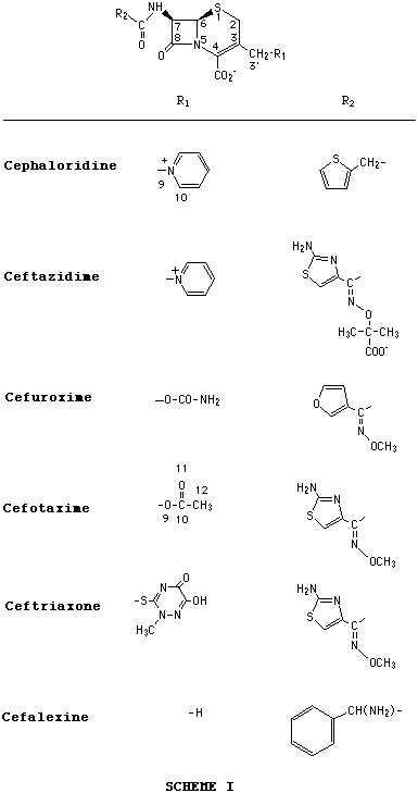
| [Molecules: None] [Related articles/posters: 040 097 098 018 044 ] |


To determine the potential influence of the substituents at C(3) and C(7) on the D2-isomerization of cephalosporins we carried out a kinetic study of the alkaline hydrolysis of cephaloridine, cefotaxime, cephalexine, cefuroxime, ceftazidime and ceftriaxone at 37 °C (Figure 1). Whenever possible, the reaction products were isolated by lyophilization to determine their structure unequivocally.
A theoretical study was performed using the AM1 molecular orbital method in order to provide support to the experimental facts. Three pairs of isomers differing in the substituent at the C(3) position were examined. An analysis of the relative thermodynamic stability of D3- and D2-isomer pairs of cephalosporins as reflected by calculated heats of formation and the influence of the C(3) substituent on the isomerization are presented.
D2-cephaloridine: 3.92 (2H, m, thienyl-CH2-CO), 4.57 [1H, s, H-C(4)], 5.31 [2H, s, C(6)-H and C(7)-H], 5.34, 5.51 (2H, AB, J=14.9 Hz, C(3)-CH2), 6.81 (1H, s, C(2)-H), 7.05 (2H, m, thienyl), 7.38 (1H, m, thienyl), 8.08 (2H, m, pyridinio), 8.58 (1H, m, pyridinio), and 8.86 (2H, d, J= 6.2 Hz, pyridinio).
D2-ceftazidime: 1.46 (s, Me2C), 4.57 [1H, s, H-C(4)], 5.43 (1H, d, J= 3.9 Hz, C(6)-H), 5.55 (1H, d, J= 3.9 Hz, C(7)-H), 5.37, 5.53 (2H, AB, J=14.6 Hz, C(3)-CH2), 6.87 (1H, s, C(2)-H), 7.02 (1H, s, C(5')-H), 8.08 (2H, m, pyridinio), 8.58 (1H, m, pyridinio), and 8.87 (2H, d, J= 6.2 Hz, pyridinio).
Theoretical studies were performed using the AM1 molecular orbital method [5] with the AMPAC 5.0 program package [6]. Calculations were carried out on a Silicon Graphics Iris Indigo XZ4000.
The D3-isomers for cephaloridine, cefotaxime and cephalexine were generated using the AMPAC 5.0 interface and the other structures (D2-isomers and intermediates of reaction) were obtained from the initial structures of the D3-isomer.
All the structures were thoroughly optimized with the Broyden-Fletcher-Goldfarb-Shanno method [7-11].
The lateral chain at position C(7) seems to have no influence on the isomerization process (as discussed below), for that reason such chains were reduced to the group CH3-CO-HN- in the studied compounds, lowering the calculation time. However, the chain at C(3) position is vital and for that reason is required in a conformational analysis of such chains in all the reactives, products and intermediates.
The method used consisted of generating a potential surface as a function of the variation of the dihedral angles C10N9-C3'C3 and N9C3'-C3C2 for cephaloridine and of the dihedral O11C10-O9C3', C10O9-C3'C3 and O9C3'-C3C2 for cefotaxime, with a step of 30°.
We have studied the alkaline hydrolysis (pH 10.5, 37 °C) of several cephalosporins by HPLC and 1H NMR. The first technique is accurate, and does not require pure compounds, however, the method does not indicate which functional group is reacting and requires an independent product study. 1H NMR lets us observe the changes in the molecule during alkaline hydrolysis.
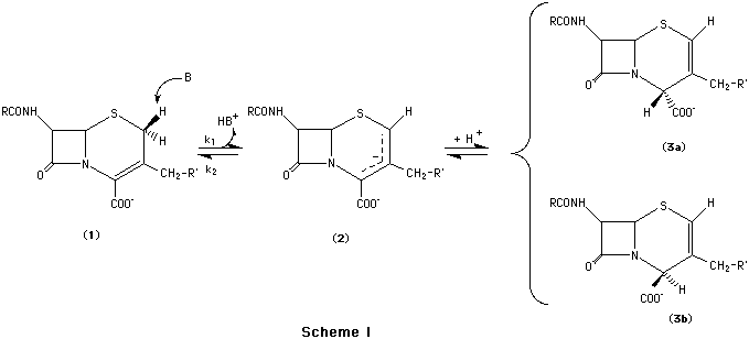
The basic hydrolysis of cephalosporins proceeds primarily via the attack of the OH- ion on the b-lactam carbonyl group, which cleaves the ring [12-13]. In cephaloridine and ceftazidime we have detected another reaction in parallel with the hydrolysis of the b-lactam ring, viz. isomerization of the double bond in the dihydrothiazine ring of the cephalosporin. In both cephalosporins we were able to isolate the D2-isomers. The presence of the D2-isomer was not detected for any of the other cephalosporins studied.
The isomerization mechanism has been widely studied [14] and of all the mechanisms suggested, the proton abstraction at C(2) mediated by a base or enzyme seems to be the most likely mechanism.
Scheme 1 shows this process of abstraction of the proton at C(2) in the D3-isomer, formation of a carbanion (2) stabilized by resonance and subsequent acceptance of the proton, partly by the C(2), regenerating the D3-isomer, or more so by the C(4) leading to the D2-isomer.
The kinetic constants obtained for the reversible base-catalysed isomerization are listed in Table 1. k1 values for cephaloridine and ceftazidime are almost identical, suggesting that the substituent at C(7) has no influence on the isomerization process.
Table 2 shows the heats of formation of structures 1, 2 and 3 for cephaloridine, cephalexine and cefotaxime. These values fail to provide a thermodynamic reason for the D3 - D2 isomerization in these antibiotics. According to the described mechanism a closer examination of the carbanion (2) is required.
Table 2 shows clearly that the intermediate 2 obtained with the cephaloridine, compound with a electron withdrawing group at C(3), is stabilized -16 kcal mol-1 (1 cal = 4.184 J) with respect to the D2-isomer. While in the cephalexine and cefotaxime, with electron donating groups, the carbanion is not stabilized with respect to the D3-isomer (+60 kcal mol-1 and +50 kcal mol-1, respectively). This result explains the different behaviour of these cephalosporins about the D2-isomerization.
The D3-D2 isomerization introduces a new asymmetric centre at C(4). Morin et al. [15] reported that only the D2 (4 a-COO-) isomer (3a) is produced. The heats of formation of both isomers [4-a-COO- (3a) and 4 b-COO- (3b)] are very similar and the 3a isomer is not always more stable than 3b. Solvation effects can affect these results and as we can see, the AMSOL method [16] predicts the 3a isomer as the most stable. However, as the difference in the heats of formation is very small, we should take into account the energetic barriers to interpret the experimental results.
In studying the double-bond isomerization of cephalosporin esters, Saab et al. [2] obtained k1 and k2 values of 0.69 and 0.32 h-1, respectively, for the methyl ester of cefazolin at pH 7.4, 40 °C and I= 0.3 M. These values are much greater than those obtained for cephaloridine and ceftazidime. Morin et al. [15] suggested that the equilibrium between D3- and D2-isomer is shifted to D2- if there are bulky groups at C(3'), in order to minimize steric repulsions. Ceftriaxone has a very bulky group at C(3'), but no D2-isomer was formed on hydrolysis, because the electron-withdrawing effect of such groups is not sufficient to promote the abstraction of a C(2)-H.
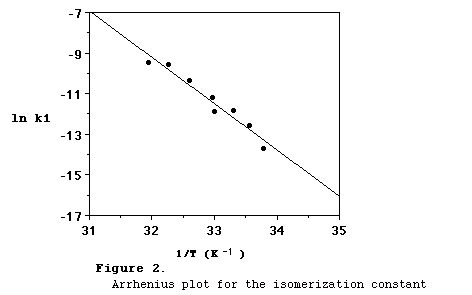
The effect of temperature on the D2-isomerization of cephaloridine was determined by measuring this rate at different temperatures (between 23 and 41 °C) in a carbonate buffer solution (pD 10.5). The pseudo-first-order rate constant was fitted to the Arrhenius equation, k= Ae-Ea/RT, by least-squares regression. The Arrhenius plot is shown in Figure 2. k1 has an energy of activation of 45.3 kcal mol-1. However, the heat of ionization of water was included in the energy of activation. By assuming such a heat to be 13.0 kcal mol-1 [17], the actual energy of activation is 32.3 kcal mol-1.

Conclusions
These results show clearly that the presence of electron donating groups does
not allow stabilization, or at least large stabilization, of the
intermediate with respect to the isomer so the formation
of the D2-isomer in cephalosporins containing an
unesterified carboxyl group is unlikely.
On the other hand, in those cephalosporins with an electron withdrawing group at C(3'), as the pyridinio ion, the negative charge on the carbanion is diminished and hence the isomerization of the double bond is favoured.
