Key words: molecular modeling/serpins/HSP47/protein C inhibitor
In order to understand the underlying mechanism of HSP47-assisted formation of procollagen triple helix, knowledge about the three dimensional structure of this protein is essential. However, there is no X-ray crystal structure of HSP47 currently available which led us to consider an alternative molecular modeling approach to obtaining structural information with which to understand structure/function relationships in this unique molecular chaperone. Molecular modeling appeared to be a valid approach to obtaining realistic structural information since in addition to being a functional molecular chaperone, HSP47 is known (9, 10, 12) to be a member of the serpin (serine protease inhibitor) superfamily of proteins which is one of the most widely and best structurally characterised protein families. There are at least 30 serpin proteins, including HSP47, which have been identified on the basis of their primary amino acid sequences (12, 13) and where X-ray crystal structure information is to hand the three dimensional structures are surprisingly consistent (13, 14). As a result, it is not unreasonable that HSP47 should adopt a similar protein fold and three-dimensional structure. Not all these serpins are functional serine protease inhibitors, but many do contain a ~ 28 amino residue loop (serpin loop) which is responsible for specific protease inhibition. Binding of this loop to a specific serine protease is followed by eventual dissociation of the serpin in a cleaved form where the amino acids to the N- and C-terminal sides of the cleaved, scissile peptide bond (i.e., respectively the P1 and P1' amino acid residues according to the Pn nomenclature of Schechter & Berger [15]) become separated by about 70 Å. Indeed, many of X-ray crystal structures of serpins which have been determined (14) are of such proteolytically cleaved serpins. Exceptions to this are the recent X-ray crystal structures of uncleaved ovalbumin (16), antichymotrypsin (ACHY) (17), plasminogen activator inhibitor-1 (PAI-1) (18) and antithrombin III (AT III) (19, 20). These recent structures have revealed that uncleaved serpins may adopt one of two main conformations. In one, the uncleaved serpin loop projects out of the protein in a conformation accessible to a serine protease active site. This inhibitory state is typified by a conformation observed in the X-ray crystal structure of AT III (19, 20). In the other, the serpin loop does not protrude, but is rendered inaccessible to a serine protease active site. This so called latent, non-inhibitory state is typified by the X-ray crystal structure of PAI-1 (18) and an alternative conformation of AT III (19).
As a result of this wealth of structural data and consistency within the serpin superfamily, several studies have been published (21, 22, 23) in which molecular modeling techniques have been used to successfully predict the structures of serpin proteins, for which no X-ray crystal structures were then available, using the X-ray crystal structure coordinates of other serpin family members. Therefore in the light of this literature precedent, the following paper outlines the application of molecular modeling, including the homology model approach, to derive appropriate three-dimensional structural information on serpin/molecular chaperone HSP47. Recently, the gene for mature recombinant mouse HSP47 (mrmHSP47) was cloned and over expressed in Escherichia coli (9), therefore we elected to model this protein specifically.
| Latent state hPCI model | Inhibitory state hPCI model | ||
|---|---|---|---|
| Residue range | R.M.S. deviation (Å) | Residue range | R.M.S. deviation (Å) |
| Pre-serpin loop region (10-356) | 0.11 | Pre-serpin loop region (10-345) | 0.08 |
| Post-Serpin loop region (368-391) | 0.26 | Post-Serpin loop region (362-391) | 0.12 |
| R.M.S. deviation (�) | |||||||||||||||||||||||||||||||||||||||||||||||||||||||||||||||||||||||||||||||||||||
|---|---|---|---|---|---|---|---|---|---|---|---|---|---|---|---|---|---|---|---|---|---|---|---|---|---|---|---|---|---|---|---|---|---|---|---|---|---|---|---|---|---|---|---|---|---|---|---|---|---|---|---|---|---|---|---|---|---|---|---|---|---|---|---|---|---|---|---|---|---|---|---|---|---|---|---|---|---|---|---|---|---|---|---|---|---|
hPCI structure element 1| Residue range in mrmHSP47 | Latent state
mrmHSP47 | Inhibitory state mrmHSP47 | Helices | A | 1-7 | 2.1 | 0.8 | B | 12-26 | 1.4 | 1 | C | 21-42 | 1.4 | 0.9 | D | 53-67 | 1.2 | 0.9 | E | 69-78 | 1.2 | 0.8 | F | 87-103 | 1.2 | 0.8 | G | 126-136 | 1.3 | 0.9 | H | 148-163 | 2 | 1.1 | I | 260-279 | 1.2 | 1.3 | Sheet A | 111-120 | 1.2 | 1.1 | 137-142 | 1.3 | 1 | 180-192 | 2 | 0.9 | 294-296 | 1 | 1.1 | 329-339 | 2.1 | 0.9 | 345-358 | 2.4 | 0.9 | Sheet B | 49-52 | 1.8 | 0.9 | 214-229 | 1.3 | 1.1 | 235-243 | 1.3 | 1.1 | 246-254 | 1.3 | 1.1 | 371-373 | 1.8 | 1.2 | 382-388 | 2 | 1.1 | Sheet C | 283-289 | 2.1 | 1.6 | |
Inhibitory and latent state models of mrmHSP47 were generated from the corresponding models of hPCI (see above) by means of homology modeling. By using the mrmHSP47/hPCI sequence alignment (Figure 1), alpha-carbon backbone coordinates of either inhibitory or latent state models of hPCI were easily assigned to corresponding residues of mrmHSP47. Thereafter, mrmHSP47 amino acid side chains were set, using the idealised geometries of CHARMm (27), and then energy minimisation protocols were followed to generate the desired inhibitory (Figure 3a) and latent (Figure 3b) state homology models of mrmHSP47. Both inhibitory and latent state homology models of mrmHSP47 were superimposed on the corresponding parent models of hPCI and the R.M.S. deviations between the major secondary structure elements recorded (Table 2).
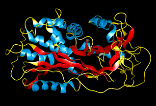
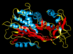
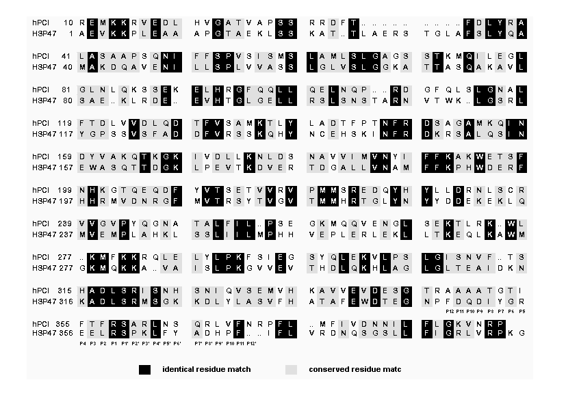
The only currently available X-ray crystal structure of hPCI (2pai, Brookhaven Protein Data Bank) is of a cleaved structure (Figure 2a) in which the peptide bond between the P1 and P1' residues (Arg358 and Ser359) (Figure 1) is proteolytically hydrolysed leaving these residues separated in space by the expected distance of 70 Å. Therefore a necessary first step in the mrmHSP47 modeling procedure was to model the uncleaved hPCI. In line with the fact that both inhibitory and latent state conformations had been observed in the recently solved X-ray crystal structures of uncleaved serpins (33), both inhibitory and latent states of hPCI were also modeled. The modeling procedure adopted to generate the inhibitory state of hPCI was based upon a previous procedure used to model the inhibitory state of alpha1-PI from the known X ray crystal structure of the cleaved form (21). The features of the resulting structure, in particular the conformation adopted by the serpin loop (yellow strand, Figure 2b), agree well with the inhibitory structure of AT III (19, 20). Also, measurements of the R.M.S. deviations (Table 1) demonstrate that the inhibitory state model of hPCI superimposes on the X-ray crystal structure of cleaved hPCI with little notable structural difference. The modeling procedure used to obtain the latent, non-inhibitory form of hPCI was designed after close inspection of the X-ray crystal structure of latent, non-inhibitory PAI-1 (18). As a result, the conformation of the serpin loop (red strand, Figure 2c) in the model is very similar to the conformation displayed in the X-ray crystal structure of latent PAI-1 (18). Once more, the latent state model of hPCI superimposes very closely on the X-ray crystal structure of cleaved hPCI (Table 1).
The approach used to model the inhibitory and latent states of mrmHSP47 was drawn from an original homology modeling procedure devised to model the inhibitory state of ACHY (23) on the refined atomic coordinates of ovalbumin (16). Homology modeling is an appropriate and effective method for determining the three-dimensional structure of a protein provided that the three-dimensional structure of a protein related by at least 25 % amino acid sequence identity is available as a template (34). Clearly hPCI and mrmHSP47 satisfied this requirement (Figure 1) thereby justifying this approach to modeling the inhibitory and latent states of mrmHSP47. The resulting homology models of inhibitory (Figure 3a) and latent (Figure 3b) states of mrmHSP47 were found to superimpose well on their respective parent models of hPCI as shown by the data in Table 2 which reports the R.M.S. deviations of individual secondary structure elements between superimposed mrmHSP47 and hPCI model structures. Generally speaking, R.M.S. deviations of <= 2.0 Å are indicative of close superposition. Table 2 shows that the R.M.S. deviations between the superimposed latent models of HSP47 and hPCI were generally larger than those between inhibitory models, especially in the region of beta-sheet A.
Whilst both latent and inhibitory states of mrmHSP47 may exist, protease inhibitor assays were carried out with purified mrmHSP47 to determine which state or conformation was more likely to exist at physiological pH-values (pH 7.5 to 7.8). The close structural relationship between hPCI and mrmHSP47, particularly the identical P1 and P1' residues (Figure 1), suggested that mrmHSP47 would behave as a functional serine protease inhibitor with the same specificity against the serine protease thrombin as hPCI (35, 36). However, in a series of standard assays (pH 7.5-8.0), mrmHSP47 not only failed to inhibit thrombin but also the serine proteases elastase, trypsin, kallikrein and tonin. Therefore the latent, non-inhibitory state of mrmHSP47 appears to predominate at standard physiological pH-values. Circular dichroism spectroscopy of mrmHSP47 at neutral pH (Miller, unpublished data) revealed that the protein contains approx. 25 % alpha-helix and 36 % beta-sheet. Gratifyingly, the latent state model of mrmHSP47 was estimated to contain approx. 26% alpha-helix and approx. 33% beta-sheet which agrees very well with the percentage levels of secondary structure experimentally determined.
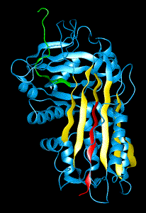
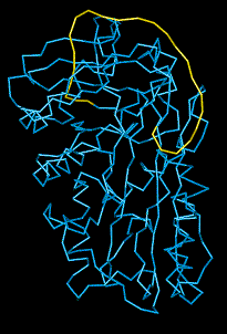
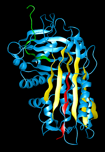
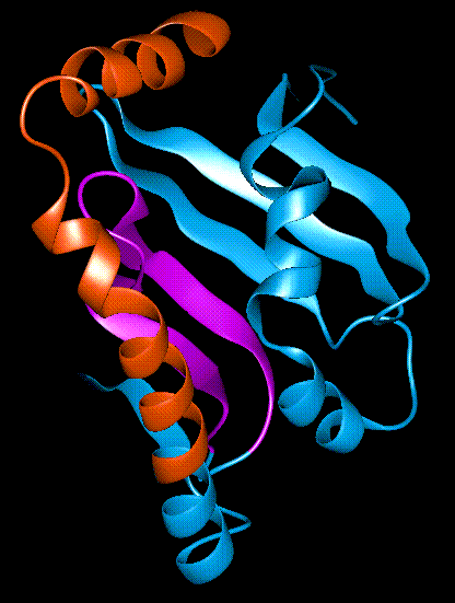
The latent state of mrmHSP47 possesses a long, deep cleft (seen in side view [Figure 3b] and top view [Figure 3c]) which appears to offer an explanation for the ability of mrmHSP47 to bind specifically and tightly to procollagen and collagen (Types I to V) peptides at standard physiological pH (11, 28, 37). The base of this cleft is formed by beta-sheet B with sides formed by helices hA and hG/hH (the helix and sheet nomenclature is derived from the Huber & Carrell labelling system [13]). Helices hA, hG/hH project hydrophilic amino acid residue side chains in towards the cleft whilst beta-sheet B projects hydrophobic amino acid residue side chains up from the bottom. This cleft is a very promising procollagen/collagen peptide binding groove for several reasons. Firstly, two deletion mutants of mrmHSP47, NΔ1 and CΔ3, have been prepared (Nagata, unpublished data) which were unable to bind collagen. NΔ1 is missing the first N-terminal 32 amino acids (Ala1-Leu32; shown in orange in Figure 3c) and CDelta 3 the last C-terminal 34 amino acids (Ala366-Leu400 or beta-strands s4B and s5B; shown in purple in Figure 3c). In both cases, the deleted peptide lengths comprise substantial parts of the putative procollagen/collagen binding groove. Secondly, this putative binding groove also bears reasonable similarity to the peptide binding regions of human class I histocompatibility glycoprotein HLA-Aw68 (38) and the human class II histocompatibility protein HLA-DR1(39). Finally, the corresponding region in hPCI has already been found to have a binding interaction with heparin (35, 40).
In conclusion, until the three-dimensional structure of mrmHSP47 is determined and refined at atomic resolution, the homology models of mrmHSP47 (Figure 3) serve as the next best structural reference for understanding structure/function relationships in serpin/molecular chaperone HSP47. This is the first time that a tandem-modeling procedure (i.e., using molecular models of an uncleaved serpin as a template for homology modeling) has been used to model the three-dimensional structure of a serpin.
2. Gething, M.-J., and Sambrook, J. (1992) Nature 355, 33-45.
3. Stress Proteins in Biology and Medicine (1990) (Morimoto, R. I., Tissieres, A., and Georgopoulos, C., Eds.), Cold Spring Harbor Laboratory Press, NY
4. Stress Proteins: Induction and Function (1990) (Schlesinger, M. J., Santoro, G., and Garaci, E., Eds.), Springer-Verlag, Heidelberg
5. Miller, A. D., Maghlaoui, K., Albanese, G., Kleinjan, D. A., and Smith, C. (1993) Biochem. J. 291, 139-144.
6. Hutchinson, J. P., El-Thaher, T. S. H., and Miller, A. D. (1994), Biochem. J. 302, 405-410.
7. Jain, N., Brickenden, A., Lorimer, I., Ball, E. H., and Sanwal, B. D. (1994), Biochem. J. 304, 61-68.
8. Wang, S.-Y. (1994)J. Biol. Chem. 269, 607-613.
9. Takechi, H., Hirayoshi, K., Nakai, A., Kudo, H., Saga, S., and Nagata, K. (1992), Eur. J. Biochem. 206, 323-329.
10. Hirayoshi, K., Kudo, H., Takechi, H., Nakai, A., Iwamatsu, A., Yamada, K. M., and Nagata, K. (1991) Mol. Cell. Biol. 11, 4036-4044.
11. Nakai, A., Satoh, M., Hirayoshi, K., and Nagata, K. (1992) J. Cell Biol. 117, 903-914.
12. Potempa, J., Korzus, E., and Travis, J. (1994) J. Biol. Chem. 269, 15957-15960.
13. Huber, R., and Carrell, R. W. (1989) Biochemistry 28, 8951-8966.
14. Schulze, A. J., Huber, R., Bode, W., and Engh, R. A. (1994), FEBS Lett. 344, 117-124.
15. Schechter, I., and Berger, A. (1967)Biochem. Biophys. Res. Commun. 27, 157-162.
16. Stein, P. E., Leslie, A. G. W., Finch, J. T., Turnell, W. G., McLaughlin, P. J., and Carrell, R. W. (1990) Nature 347, 99-102.
17. Wei, A., Rubin, H., Cooperman, B. S., Schechter, N., and Christianson, D. W. (1994) Nature Str. Biol. 1, 251-257.
18. Mottonen, J., Strand, A., Symersky, J., Sweet, R. M., Danley, D. E., Geoghegan, K. F., Gerard, R. D., and Goldsmith, E. J. (1992)Nature 355, 270-273.
19. Carrell, R. W., Stein, P. E., Fermi, G., and Wardell, M. R. (1994), Structure. 2, 257-270.
20. Schreuder, H. A., de Boer, B., Dijkema, R., Mulders, J., Theunissen, H. J. M., Grootenhuis, P. D. J., and Hol, W. G. J. (1994) Nature Str. Biol. 1, 48-54.
21. Engh, R. A., Wright, H. T., and Huber, R. (1990) Prot. Eng. 3, 469-477.
22. Jarvis, J. A., Munro, S. L. A., and Craik, D. J. (1992) Prot. Eng. 5, 61-67.
23. Katz, D. S., and Christianson, D. W. (1993) Prot. Eng. 6, 701-709.
24. Needleman, S. B., and Wunsch, C. D. (1970) J. Mol. Biol. 48, 443.
25. Atlas of Protein Sequence and Structure (1978) (Dayhoff, M. O., Ed.) 5(3), NBRF, Silver Spring, MD.
26. Feng, D. F., and Doolittle, R. F. (1987)J. Molec. Evol. 25, 351-360.
27. Brooks, B. R., Bruccoleri, R. E., Olafson, B. D., States, D. J., Swaminathan, S., and Karplus, M. (1983)J. Comp. Chem. 4, 187-217.
28. Saga, S., Nagata, K., Chen, W.-T., and Yamada, K. M. (1987), J. Cell Biol. 105, 517-527.
29. Warburg, O., and Christian, W. (1941)Biochim. Z. 310, 384-421.
30. Lottenburg, R., Christensen, U., Jackson, C. M., and Coleman, P. L. (1981), Meth. Enzymol. 80, 341-361.
31. Castillo, M. J., Nakajima, K., Zimmerman, M., and Powers, J. C. (1979), Anal. Biochem. 99, 53-64.
32. El-Thaher, T. S. H. & Bailey, G. S. (1993) Int. J. Peptide Protein Res. 41, 196-200.
33. Goldsmith, E. J., and Mottonen, J. (1994) Structure 2, 241-244.
34. Blundell, T. L., Sibanda, B. L., Sternberg, M. J. E., and Thornton, J. M. (1987), Nature 326, 347-352.
35. Whinna, H. C., and Church, F. C. (1993) J. Prot. Chem. 12, 677-688.
36. Carrell, R. W., and Boswell, D. R. (1986) in Serpins: The superfamily of plasma serine proteinase inhibitors. Proteinase Inhibitors , pp 403-420, Elsevier Science BV.
37. Natsume, T., Koide, T., Yokota, S., Hirayoshi, K., and Nagata, K. (1994), J. Biol. Chem. 269, 31224-31228.
38. Silver, M. L., Guo, H.-C., Strominger, J. L., and Wiley, D. C. (1992), Nature 360, 367-369.
39. Stern, L. J. , Brown J. H., Jardetsky, T. S., Gorga, J. C., Urban, R. G., Strominger, J. L., and Wiley, D. C. (1994) Nature 368, 215-221.
40. Shirk, R. A., Elisen, M. G. L. M., Meijers, J. C. M., and Church, F. C. (1994), J. Biol. Chem. 269, 28690-28695.
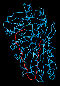
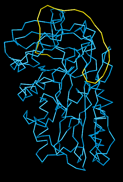
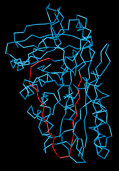
Fig. 2 a. X-ray crystal structure of human protein C inhibitor (hPCI). beta-Sheet A is shown in yellow, beta-strand s4A in red and beta-strand s1C in green. Huber and Carrell (13) nomenclature applies. 2b. Inhibitory state model of hPCI. Modeled serpin loop is shown in yellow. 2c. Latent state model of hPCI. Modeled serpin loop is shown in red.
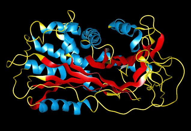
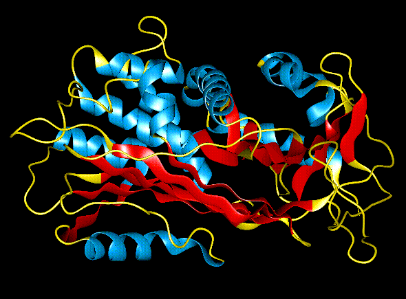
Fig. 3 a. Side view of inhibitory state homology model of mature recombinant mouse HSP47 (mrmHSP47). alpha-Helices are coloured blue, beta-strands red and loops yellow. 3b. Side view of latent state homology model of mrmHSP47. alpha-Helices are coloured blue, beta-strands red and loops yellow. 3c. Close-up, top view of the putative procollagen/collagen binding groove. The groove has been coloured coloured to show the peptide lengths of mrmHSP47 deleted in mutant NDelta 1, (orange segment) and CDelta 3 (purple segment).